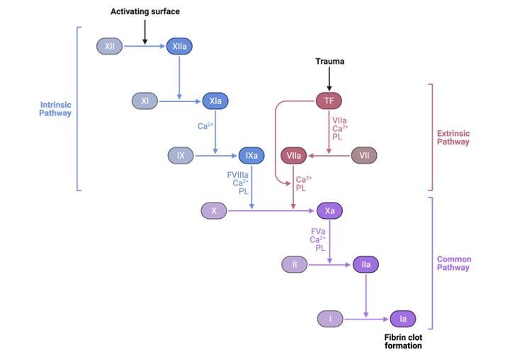
The blood first transforms from a liquid to a gel. At least 12 chemicals known as clotting factors or tissue factors are involved in a series of chemical events that result in the fibrin meshwork inside the blood. Each clotting factor has a distinct purpose. The key variables in the coagulation cascade’s result are prothrombin, thrombin, and fibrinogen.
The liver produces prothrombin and fibrinogens, which are proteins that are deposited in the blood. When blood arteries are injured, adjacent platelets are prompted to produce a chemical called prothrombin activator, which stimulates the conversion of prothrombin, a plasma protein, into thrombin, an enzyme. Calcium ions are required for this process.
This is where the calcium metabolism that we discussed before comes into play, which is present in both vitamin K2 and chitosan because both contain calcium ions. So this is when the entire hemostatic process comes to a halt.
Thrombin aids in the conversion of fibrinogen, a soluble plasma, into long insoluble fibres or threads of fibrin, a protein. Fibrin threads form an interlocking network of fibres and the structure for the clot as they loop around the platelet plug at the injured region of the blood artery.
The net of fibres catches and retains platelets, blood cells, and other substances close to the damage site, making the clot more effective. This transient fibrin clot forms in under a minute and restricts blood flow until platelets join. As a result, there is a link between calcium ions and calcium metabolism, which becomes engaged in the process and produces the first clot.
As a result, chitosan and Vitamin K2 interact with calcium ions in this way. Chitosan chitin and vitamin K2 have a lot in common. Chitin is a glucosamine-based polymer found in the shells and walls of crustaceans, fungus, and yeast. It is made up of calcium oxide and protein units and is the primary component of crustacean exoskeletons.
In crab shells, chitin accounts approximately 50-80% of organic molecules. Shellfish chitin is treated with alkali to produce chitosan, an amino polysaccharide. Chitin and chitosan are nonstarch polysaccharides that have the potential to be considered dietary fibre components.
Vitamin K3, on the other hand, has impacts on calcium ions and bone production, as stated above. As a result, there is a link between the body’s calcium metabolism and blood clotting that requires more investigation. The research has progressed.
Every day, experts come out with fresh research remedies for the same problem. Coagulation is now carried a step further by entering the coagulation pathways, which will be discussed in more detail in relation to calcium metabolism. Hemostatic arrest bleeding, which is the fundamental function of chitosan and vitamin K2, is a commonly utilised application for blood clotting.
In the therapy of wound healing, a functional chitosan-based hydrogel containing vitamin K2 can be used as a wound dressing and drug delivery system. Function active wound dressings are supposed to maintain a moist wound environment, protect against secondary infections, eliminate wound exudate, speed tissue regeneration, and increase wound healing efficiency.
Biodegradable, biocompatible, non-toxic, antibacterial, biologically adhesive, biological activity, and hemostatic properties make chitosan-based hydrogels excellent materials for aiding wound healing.
Chitosan-based hydrogels containing vitamin K2 have been shown to enhance wound healing at various phases of healing and to relieve variables that impede wound healing, such as excessive inflammatory response and persistent wound infection.
Because of the unique biological characteristics of chitosan-based hydrogel containing vitamin k2, it may be used as a wound dressing as well as a drug delivery system (DDS) to administer antibacterial agents, growth factors, stem cells, and other substances that can help speed wound healing.
By altering or combining with other polymers and containing various types of active ingredients, chitosan-based hydrogels can improve the efficiency of wound healing for a variety of wounds. Let’s take a deeper look at how chitosan-based hydrogels containing vitamin K2 may be used to improve wound healing and drug delivery systems.