What is Alimentary Canal?
o Alimentary canal, also known as gastro-intestinal tract is where the complete human digestion takes place starts from mouth and ends in the anus.
o The whole alimentary canal is divided into different compartments that is buccal cavity, esophagus, stomach, large intestine along with rectum and last part is anus.
o The process of digestion is ingestion followed by digestion to assimilation of the digested food then egestion of the waste product.
Layers of Alimentary Canal
o The alimentary canal has different layers that is uniform throughout the canal.
o The alimentary canal layers are mucosa, submucosa, muscular layer and serosa with adventitia.
I. Mucosa
o It is the mucus membrane as for the production of mucus is characteristics of the epithelium.
o It has layer of epithelium that is in direct contact with the indigested food and the loose connective tissue, lamina propria along with the smooth muscle layer called muscularis mucosa.
o The stratified squamous epithelium of mucosa layer is present in the mouth, pharynx, oesophagus and anal canal. Whereas the intestines and stomach have columnar epithelium.
o Epithelial cells have average lifespan of couple days to weeks.
o Renewal of the epithelial cells in preserving the alimentary canal condition from the wear tear from the food stuffs.
o Lamina propria consists of loose connective tissue along with the blood and lymphatic vessels that helps in the transportation of absorbed nutrients to different parts.
o Muscularis mucosa has thin layer of smooth muscle as this layer is in continuous tension. This layer increases surface area of the digestion and absorption.
Diagram Representing Layers of Alimentary Canal
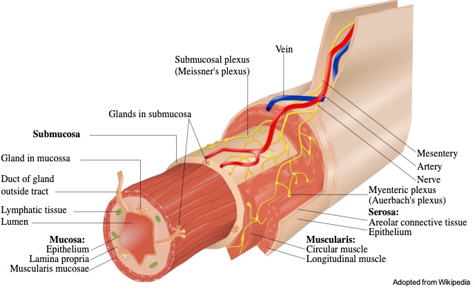
II. Sub-Mucosa
o Submucosa consists of the thick layer of connective tissue lying beneath the mucosa. It also has blood vessels, lymphatic and nerves branching.
III. Muscular Layer
o There are two layers, inner is a circular smooth muscle whereas outer layer is longitudinal smooth muscles. These are used for the peristalsis movement to pass the food through the gut.
III. Serosa and Adventitia
o The final layers of alimentary canal have loose connective tissue and coated with mucus to prevent damage due to friction between intestine and other tissue.
o Regions within peritoneum is serosa that are small intestine, stomach, caecum, appendix, rectum, sigmoid colon, transverse colon and regions behind peritoneum have adventitia which includes stomach, distal duodenum, oral cavity, oesophagus, pylorus of stomach, anal canal, ascending and descending canal.
Alimentary Canal Diagram

Alimentary Canal Structure and Function
I. Buccal Cavity
o Buccal cavity constitutes the teeth, tongue and saliva. The process of intaking of food into the mouth is called ingestion.
o Teeth and tongue help in the breakdown of the food completely and mixing it with saliva. Saliva helps the breakdown of starch into sugars.
o Tongue also helps in the process of swallowing of food and passing it to the esophagus/ food pipe.
II. Esophagus
o Buccal cavity is followed by pharynx, a short passage common for both air and food.
o Trachea and esophagus open into pharynx which has a flap like opening called epiglottis that prevents the food from entering the food into the glottis (opening of wind pipe) while swallowing.
o Swallowed food is passed into the food pipe and through the thorax (region between the neck and abdomen).
o Mucus present in the saliva is mixed with the food that helps in the adhering the food particles together forming bolus.
o Bolus is passed on with the help of the muscular contraction waves called peristalsis.
o The food from the esophagus moves to the stomach.
o Gastro – esophageal sphincter helps controlling the passage of food into the stomach.
III. Stomach
o Stomach secretes many digestive juices along with hydrochloric acid and mucus.
o Mucus acts as protective covering to the stomach lining from the hydrochloric acid whereas the digestive juices help in breakdown of proteins.
o Hydrochloric acid in addition to the breakdown of food, it kills the bacteria that enters the stomach.
o The stomach is a J shaped organ located in the upper left corner of the abdominal cavity.
o Stomach is known to have three parts namely, cardiac region opens to the esophageal opening, fundic region, where the food is stored and breaks down and pyloric region that opens to the small intestine.
IV. Small Intestine
o Stomach is followed by the small intestine, a C shaped organ consisting of three parts, duodenum, jejunum (long coiled middle part) and ileum (highly coiled).
o The opening of small intestine from stomach is controlled by the pyloric sphincter.
o Small intestine is 5m coiled but is highly coiled and receives digestive enzymes from pancreas (insulin glucagon) and liver (bile) and itself produces digestive secretion.
o The broken food particles mixed with the enzymes is moves through the walls of small intestines.
o The presence of finger like projection on the walls of intestines helps in the absorption of the digested food that is passed into blood vessels of the small intestines as villi forms the network of capillaries in addition to large lymph vessel.
o The absorbed food particles from the walls of intestine are passed onto different parts of the body is called assimilation takes place.
o The undigested food is passed into large intestine.
V. Large Intestine and Rectum
o When compared to small intestine, large intestine is wider and shorter than small intestine with length of 1.5m.
o Large intestine helps in further digestion of the undigested food and absorption of water.
o Large intestine has different parts called as caecum, colon (ascending colon, transverse colon, descending colon, sigmoid colon) and rectum.
o Both small and large intestine have goblet cells but large intestines have more of goblet cells.
o The goblet cells secrete mucin to form mucus in the water helps in adhering the waste and lubricating the waste to pass easily.
o The unabsorbed and undigested food is passed through caecum to the rectum.
o The ileo – caecal valve prevents the back flow of the fecal matter.
o The feces is stored in the rectum till defecation.
VI. Anus
o Anus is the gastro-intestinal tract and the last part of the canal.
o Rectum followed by the anus and the anorectal line separates rectum and anus.
o The anus is surrounded by the tissue called fascia.
o The walls of anus is formed the external sphincter and there are fluid secretion by the glands of anus to keep it moist.
You may like to read;
Human Digestive System: Diagram, Parts and Function
Mitochondria: Function, Definition, Significance, and Facts
Krebs Cycle: Definition, Diagram, Steps, and Mechanism
Breast Cancer: Symptoms, Stages, Diagnosis, and Treatment
Alimentary Canal Citations
- Total alimentary canal necrosis: infarction from oesophagus to anus. BMJ Case Rep . 2017 Aug 16;2017:bcr2017221957.
- The microstructure of buccal cavity and alimentary canal of Siganus rivulatus: Scanning electron microscope study. Microsc Res Tech . 2019 Apr;82(4):443-451.